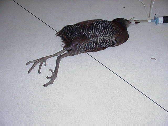

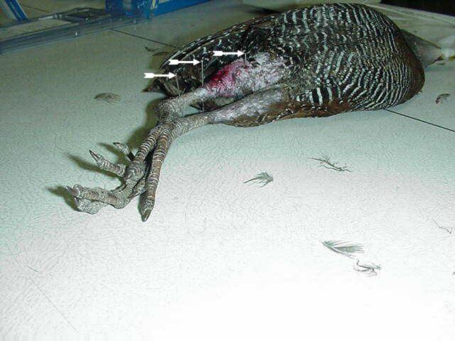
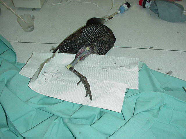
Above left and right: Physical
exam findings of right and left valgus deformity of the legs associated
with right and left tibiotarsal rotation. The patient has been induced
to a surgical plane of anesthesia with isoflurane gas delivered in 100%
oxygen via a norman elbow with a 2.0 mm non-cuffed endotracheal tube.
Non-cuffed endotracheal tubes are recommended for avian species, as cuffed
tubes tend to result in tracheal necrosis in a relatively high percentage
of cases. Respiration is being monitored with an in-line respirometer
(right figure, top left corner).
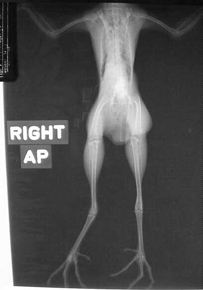
Above left:
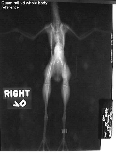
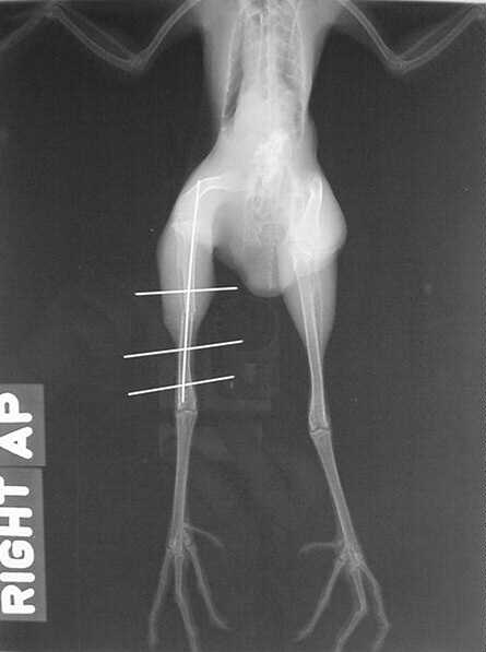
Above right:
VD view showing curled toes and tibiotarsal
rotation.
Normal Guam rail reference VD view for
comparison.
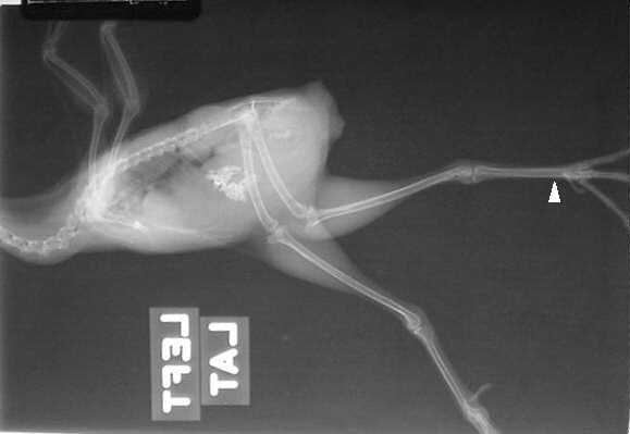
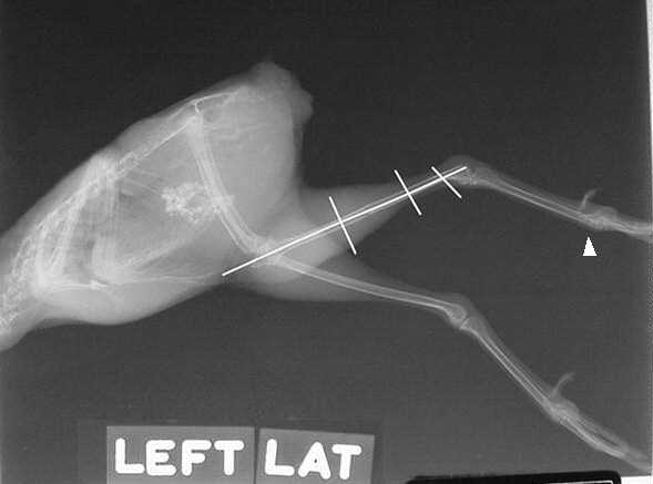
Above: Lateral radiograph
showing virtually no angular deformity of the tibiotarsii other than rotation.
Note the unusual appearance of the right foot (upper limb) as a result
of the valgus deformity. This "AP" view should be seen only in the
VD view (arrow).
Treatment: Anesthesia was induced with isoflurane delivered through a Norman elbow via 2.0 mm uncuffed endotracheal tube. To minimize the risk of managing a "downer" bird only one limb at a time was corrected. The surgical goal was to derotate the tibiotarsii and then attempt to straighten the toes post derotation using appropriate splinting materials and tape sandals.
The right tibiotarsus was fractured at about the midshaft point by repeatedly drilling a 0.028" K wire through the diaphysis until the bone was weakened enough to be easily fractured. A three-pin, through-and-through Kirschner Ehmer apparatus was applied using 0.028" K wires. In addition, a single 0.028" K wire was placed intramedullary in the right tibiotarsus to provide better stability. Originally a four-pin through-and-throug KE was going to be applied, but the proximal pin would have interfered with normal mobility by protruding too far into the lateral abdomen, and so was eliminated from the repair. The intramedullary K wire was used to help compensate for the lack of complete stability of the repair from the lack of the proximal K wire in the through-and-through KE.
Above: Lateral radiograph
of a three-pin through-and-through 0.028" K wires placed, along with a
single intramedullary 0.028" K wire. Note that the right foot no
longer has the "AP" appearance in this view as a result of the repair attempt
(arrow).
Above: VD view of the 0.028"
K wire three-pin, through-and-through KE with a single 0.028" K wire intramedullary
pin in the right tibiotarsus.
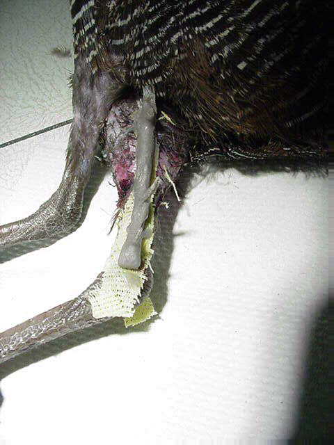
Above: Three-pin through-and-through 0.028" K wires placed into the right tibiotarsus (arrows). The K wires were placed with a small hand chuck.
Above: The three-pin KE has been stabilized laterally with a methylmethacrylate bar. The technique used was to first impale a protective strip of xeroform gauze, followed by a strip of1/8" umbilical tape over the pins. Next, the umbilical tape was impregnated with methacrylate. The result is a light weight and sturdy KE bar that is easy to apply with minimal distraction of the pins (see image below):
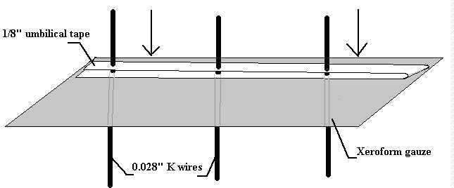
Above: Xeroform gauze (gray
thick rectangle) is forced over the pins (black lines), followed by umbilical
tape (white thinner rectanges). Once the umbilical tape is in place
methacrylate is painted liberally onto the umbilical tape to form a lightweight
KE bar.
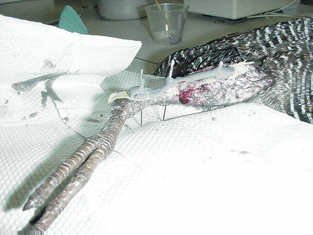
Above: Once the methacrylate
bar on the lateral aspect has hardened, the bird is turned over, and a
similar stabilizing bar is applied to the medial aspect of the right tibiotarsus
using an identical technique to that applied to the lateral stabilizing
bar.
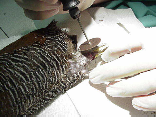
Above: Once the methacrylate on the lateral and medial stabilizing bars has set a carborundum blade or clippers are used to cut down the K wires to minimize trauma to the patient. Care must be taken when using the dremel with the carborundum blades, as the K wires can become hot enough to cause necrosis of the tissue and bone. In this case the procedure was stopped several times to allow alcohol to be dripped onto the methacrylate and the K wire to cool the apparatus.
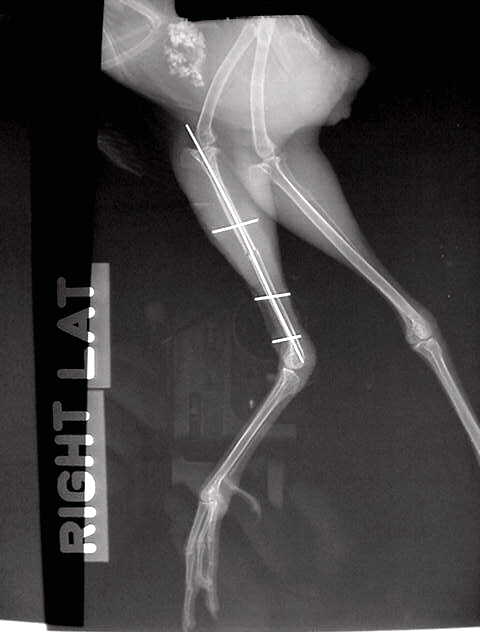
Above: Postoperative lateral
radiograph of the repair on the right tibiotarsus showing good alignment
of the proximal and distal fracture fragments.
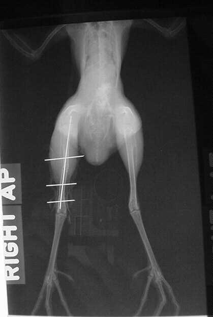
Above: Postoperative VD radiograph showing good alignment of the proximal and distal fracture fragments with improved conformation of the right tibiotarsus, despite the minimal valgus deformation of the tibiotarsus resulting from the imperfect intramedullary 0.028" K wire. When the bird recovered it was able to ambulate on the right leg, though was lame during the immediate postoperative period. Butorphanol was given at 1 mg/kg p.o. for the first postoperative day to decrease pain. Followup: The bird regained 80% of normal function, with some slight medial curling of the digits as a residual deficit, but is functionally normal.
Summary: The cause of the deformity is likely associated with excessive dietary protein. This is the fourth case of tibiotarsal deformity in Guam rail that has been encountered in the last 3 years in our colony. Like cranes, the Guam rail (and perhaps other rallidae) may be susceptible to bone deformation during growth resulting from relatively high dietary protein concentration. The repair was performed in an attempt to restore more normal function to this otherwise healthy bird.
Patrick J. Morris, D.V.M., D.A.C.Z.M.
Veterinary advisor for the Gruiformes
TAG, Rallidae*
* SSP and TAG are both managed under
the auspices of the American
Zoological Association